Ear reconstruction encompasses all operations used in the treatment of ear deformities. These conditions include microtia (congenital underdeveloped, or missing, ears), constricted ear deformities, protruding ears and acquired ear anomalies.
Autologous ear reconstruction using rib cartilage
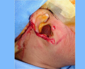
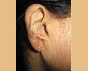
Autologous ear reconstruction requires multiple surgical stages. During the initial stage, cartilage is taken from the ribs and used to create a three-dimensional sculpture that looks like a normal ear. This is the technique used by most surgeons, and is typically performed when the patient is between 6 and 10 years of age. At this age, the opposite ear is close to adult size and the ribs will provide enough material to create a framework of adequate size.
The cartilage framework is then placed under the skin at the site of the newly placed ear, to match the opposite ear. Additional cartilage may be saved either at the rib donor site or underneath the temporal scalp for use in a later stage. Suction catheters are placed under the skin to create better definition around the framework. They are removed in one to two weeks. Children are usually admitted to the hospital for three to five days.
Several techniques have been adopted for autologous ear reconstruction, i.e. the Brent method and the Nagata method. The differences in these techniques lie in the number of stages required for ear reconstruction (between 2 and 4) and the amount of cartilage needed to make the ear framework. For the Nagata method, children should be closer to 10 years of age to make sure there is enough cartilage to take from the ribs.
Play Video about ear reconstruction
CASE 1
CASE 2
CASE 3
CASE 4
dideesh mohan
2022-07-13
Trustindex verifies that the original source of the review is Google.
നമസ്തേ
എൻ്റെ പേര് ദിദീഷ് കഴിഞ്ഞ ആഴ്ച ഞാൻ ഒരു അപകടം പറ്റി Elite ഹോസ്പിറ്റലിൽ അഡ്മിറ്റ് ആയിരുന്നു ഒപ്പം എൻ്റെ ചേട്ടനും ഉണ്ടായിരുന്നു ഡോർ കട്ട് ചെയ്യുന്നതിനിടെ കട്ടർ തെറിച്ചു കാൽപാദം പകുതി മുറിഞ്ഞ് . ചേട്ടന് കാൽമുട്ടിൽ ആയിരുന്നു ഓപറേഷൻ കഴിഞ്ഞു വീട്ടിൽ ആണ് രണ്ടാളും സുഖമായിരിക്കുന്നു. എൻ്റെ കാൽപാദം മുറിച്ചു മാറ്റും എന്ന ഒരു പേടി മാത്രേ എനിക്കുണ്ടായിരുന്നുള്ളു പക്ഷെ അങ്ങനെ ഒന്നും സംഭവിച്ചില്ല എല്ലാ സ്റ്റാഫിനും നന്ദി പറയുന്നു കാരണം വന്നപ്പോ തൊട്ട് എല്ലാവരും സമാതാനിപ്പിക്കായിരുന്നൂ എല്ലാവരോടും സ്നേഹം അറിയിക്കുന്നു പ്രത്യേകിച്ച് ഡോക്ടറോട് 🙏🏻
SHIJI RAMESH
2022-07-13
Trustindex verifies that the original source of the review is Google.
Excellent center for all cosmetic procedures...
highly recommended
Shafi Sanu
2022-06-21
Trustindex verifies that the original source of the review is Google.
The surgery is in a seamless hospital and everything is very excellent